Arteries Diagram : Anatomy And Function Of The Coronary Arteries Johns Hopkins Medicine. After receiving blood directly from the left ventricle of the heart, the. 10+ heart lung circulation diagram. The arteries' smaller branches are called arterioles and capillaries. Like maps, the various diagrams emphasize different aspects. Smartdraw includes 1000s of professional healthcare and anatomy chart templates that you can modify and make your own.
The journal promotes multidisciplinary dialogue between. After receiving blood directly from the left ventricle of the heart, the. From this trunk, several vessels arise, which go on to supply the neck. Two major coronary arteries branch off from the aorta near the point where the aorta and the left ventricle meet. The cardiovascular system consists of the heart, blood vessels, and the approximately 5 liters of blood that the blood vessels transport.
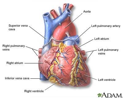
Heart Front View Medlineplus Medical Encyclopedia Image from medlineplus.gov John bavosi/science photo library/getty images. Cardiovascular system human veins arteries heart continued from cardiovascular system anatomy the heart the heart is a muscular pumping organ located medial to the lungs along the body's human body muscle diagram a fully labelled human body muscle diagram 4 fantastic large size a4 labeled human body muscular system pictures for you to print and then There are two types of them: From this trunk, several vessels arise, which go on to supply the neck. The main artery to the leg is the femoral artery.it is the continuation of the external iliac artery below the inguinal ligament. Pulmonary circulaton brings blood into contact with alveoli in the lungs for gas exchange (this is the only place in body where arteries carry o2 poor blood and veins. Smartdraw includes 1000s of professional healthcare and anatomy chart templates that you can modify and make your own. Cardiac veins then drain away the blood after it has been deoxygenated.
Learn the differences between an artery and a vein.
Carotid artery disease is caused by a buildup of plaques in arteries that deliver blood to your brain. 10+ heart lung circulation diagram. Human body artery diagram in detail. The arteries' smaller branches are called arterioles and capillaries. 14+ heart arteries diagram labeled. From this trunk, several vessels arise, which go on to supply the neck. Coronary arteries supply blood to the heart muscle. The first branch of the thyrocervical trunk is the inferior thyroid artery. Pulmonary circulaton brings blood into contact with alveoli in the lungs for gas exchange (this is the only place in body where arteries carry o2 poor blood and veins. An artery is an elastic blood vessel that transports blood away from the heart. Arteries of the lower limb thigh leg foot the main artery of the lower limb is femoral artery it is a continuation of the external iliac artery terminal branch of the abdominal aorta the arteries and veins of the leg smartdraw arteries and veins of the leg create healthcare diagrams like this example called arteries and veins of the leg in minutes with smartdraw. After receiving blood directly from the left ventricle of the heart, the. It ends at the anterior and posterior tibial arteries.
Arteries and veins of the arm. Inner body parts with their names. The coronary arteries wrap around the outside of the heart. Arteries carry blood away from the heart in two distinct pathways: 14+ heart arteries diagram labeled.
Connectivity Of The 55 Main Arteries In The Human Arterial System Download Scientific Diagram from www.researchgate.net Other arteries of the neck. Ascending aorta, aortic arch, thoracic aorta, and abdominal aorta. Cardiac veins then drain away the blood after it has been deoxygenated. The external iliac artery is a branch of the common iliac artery which is formed when the abdominal aorta bifurcates (divides into two). From this trunk, several vessels arise, which go on to supply the neck. The two exceptions are the pulmonary and the umbilical arteries, which carry deoxygenated blood to the organs that oxygenate it (lungs and placenta. Arteries are quite tough on the outside but are smooth on the inside. In the femoral triangle, the profunda femoris artery arises from the.
Plaques are clumps of cholesterol, calcium, fibrous tissue and other cellular debris that gather at microscopic injury sites within the artery.
There are two types of them: Pulmonary circulaton brings blood into contact with alveoli in the lungs for gas exchange (this is the only place in body where arteries carry o2 poor blood and veins. The tunicae intima, media, and externa. The main artery to the leg is the femoral artery.it is the continuation of the external iliac artery below the inguinal ligament. It is a continuation of the external iliac artery (terminal branch of the abdominal aorta). Other arteries of the neck. An artery (plural arteries) (from greek ἀρτηρία (artēríā) 'windpipe, artery') is a blood vessel that takes blood away from the heart to one or more parts of the body (tissues, lungs, brain etc.). In the femoral triangle, the profunda femoris artery arises from the. The main artery of the lower limb is the femoral artery. These arteries and their branches supply all parts of the heart muscle with blood. The journal promotes multidisciplinary dialogue between. The external iliac becomes the femoral artery when it crosses under the inguinal ligament and enters the femoral triangle. Coronary arteries supply blood to the heart muscle.
From this trunk, several vessels arise, which go on to supply the neck. The coronary arteries wrap around the outside of the heart. The cardiovascular system consists of the heart, blood vessels, and the approximately 5 liters of blood that the blood vessels transport. In this image, you will find external carotid artery, internal carotid artery, vertebral artery, aorta and arch, pulmonary artery, cardiac artery, thoracic aorta, celiac trunk, superior mesenteric artery, renal artery, gonadal artery, inferior mesenteric artery, common iliac artery, external iliac artery. The main artery of the lower limb is the femoral artery.
Pulmonary Artery Wikipedia from upload.wikimedia.org Next, we have the blood vessel responsible for carrying deoxygenated blood from the right side of the heart (right ventricle) to the lungs. In honor of valentine's day, we're learning about the heart and other major organs! Arteries carry blood away from the heart in two distinct pathways: The main artery to the leg is the femoral artery.it is the continuation of the external iliac artery below the inguinal ligament. John bavosi/science photo library/getty images. The heart receives its own supply of blood from the coronary arteries. Coronary arteries supply oxygenated blood to the heart muscle. In this image, you will find external carotid artery, internal carotid artery, vertebral artery, aorta and arch, pulmonary artery, cardiac artery, thoracic aorta, celiac trunk, superior mesenteric artery, renal artery, gonadal artery, inferior mesenteric artery, common iliac artery, external iliac artery.
Human body artery diagram in detail.
The main artery of the lower limb is the femoral artery. This is known as the main pulmonary artery or pulmonary trunk. A wire is moved through an artery in the leg up to the carotid artery, and a small wire tube, or stent is expanded inside a narrowing of the carotid artery. Cardiovascular system human veins arteries heart continued from cardiovascular system anatomy the heart the heart is a muscular pumping organ located medial to the lungs along the body's human body muscle diagram a fully labelled human body muscle diagram 4 fantastic large size a4 labeled human body muscular system pictures for you to print and then In honor of valentine's day, we're learning about the heart and other major organs! Arteries of the lower limb thigh leg foot the main artery of the lower limb is femoral artery it is a continuation of the external iliac artery terminal branch of the abdominal aorta the arteries and veins of the leg smartdraw arteries and veins of the leg create healthcare diagrams like this example called arteries and veins of the leg in minutes with smartdraw. Next, we have the blood vessel responsible for carrying deoxygenated blood from the right side of the heart (right ventricle) to the lungs. The aorta branches into a network of smaller arteries that extend throughout the body. This is an excellent human heart diagram which uses different colors to show different parts and also labels a number… Coronary arteries supply oxygenated blood to the heart muscle. Arteries and veins of the arm. Two major coronary arteries branch off from the aorta near the point where the aorta and the left ventricle meet. Learn the differences between an artery and a vein.